this post was submitted on 14 Jan 2025
641 points (100.0% liked)
pics
19813 readers
810 users here now
Rules:
1.. Please mark original photos with [OC] in the title if you're the photographer
2..Pictures containing a politician from any country or planet are prohibited, this is a community voted on rule.
3.. Image must be a photograph, no AI or digital art.
4.. No NSFW/Cosplay/Spam/Trolling images.
5.. Be civil. No racism or bigotry.
Photo of the Week Rule(s):
1.. On Fridays, the most upvoted original, marked [OC], photo posted between Friday and Thursday will be the next week's banner and featured photo.
2.. The weekly photos will be saved for an end of the year run off.
Instance-wide rules always apply. https://mastodon.world/about
founded 2 years ago
MODERATORS
you are viewing a single comment's thread
view the rest of the comments
view the rest of the comments


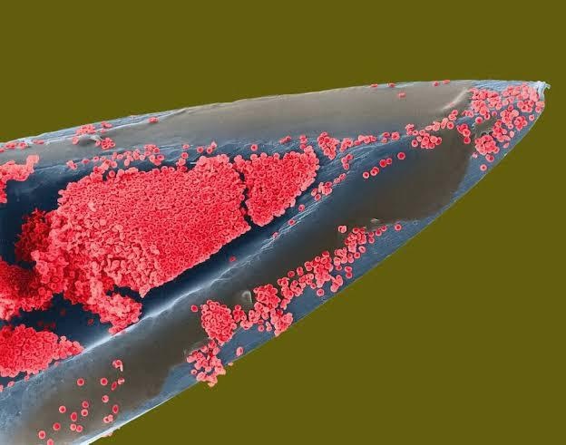
When I segmented 3D MRI and CT scan images before I used the contrast borders for help a lot. There were some algorithms for finding edges that you could tune by setting search radiuses and thresholds. There was also an option of growing an area by a certain amount of pixels outward, and then threshholding the result back down to only the brighter parts, that kind of thing. You had to be a little clever about how you'd combine it. And ultimately, sometimes I just had to add and subtract a few points manually.
Segmenting is more assigning areas to distinct objects (separating bones from the rest in my case), but you could totally use it as a basis for coloring, so I assume the process is similar here.